Caption
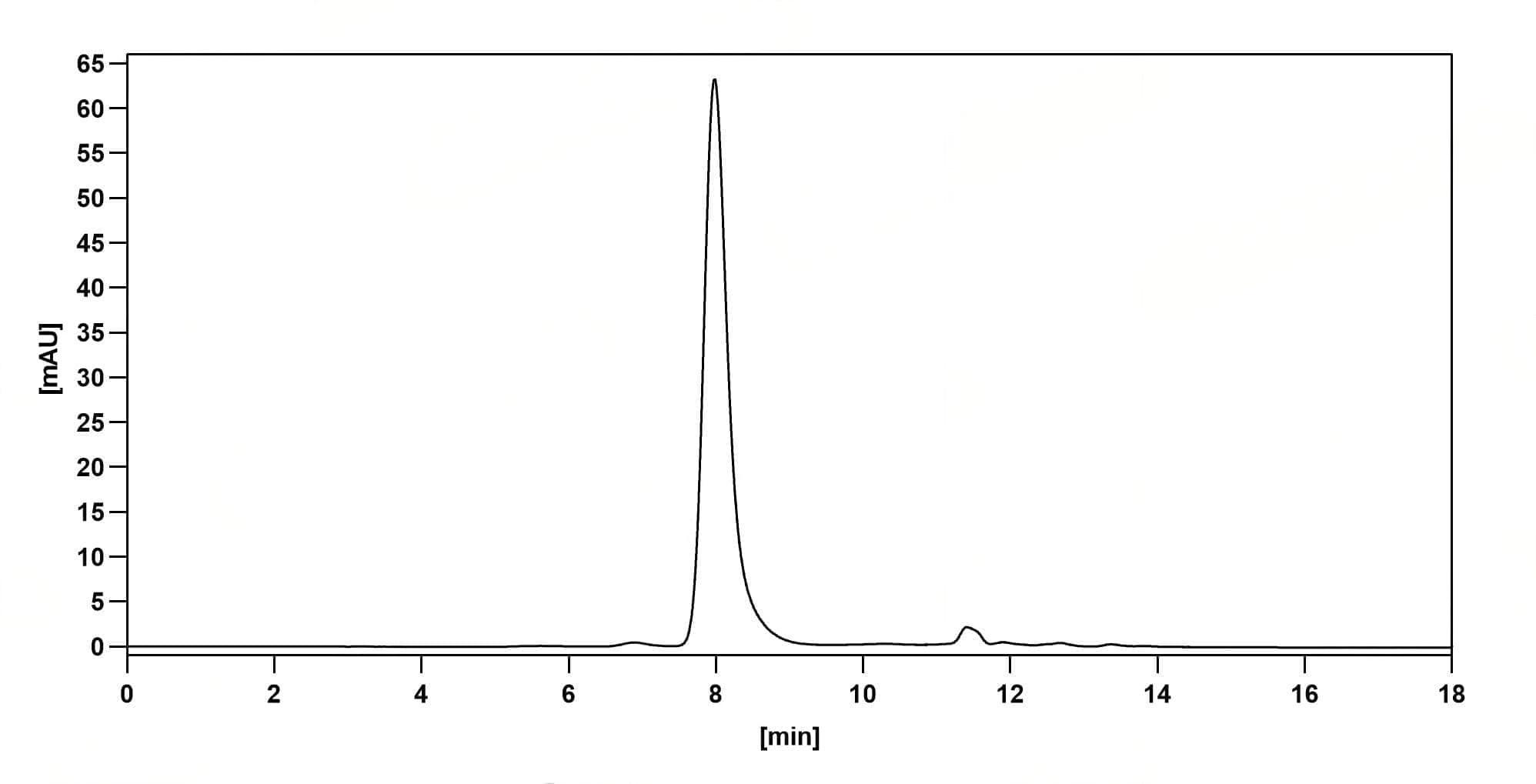
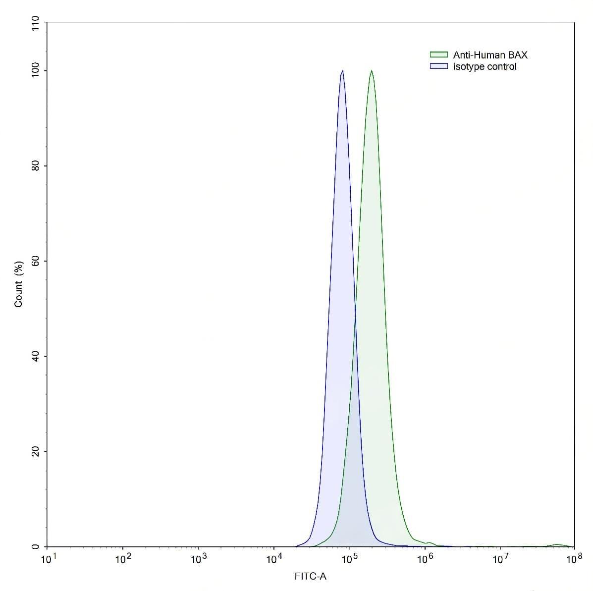
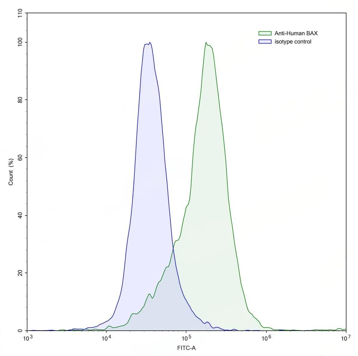
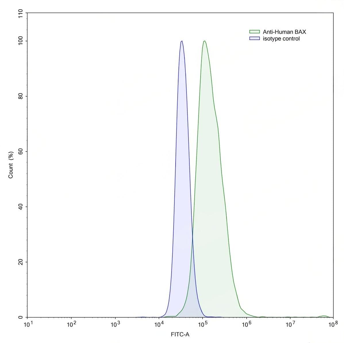
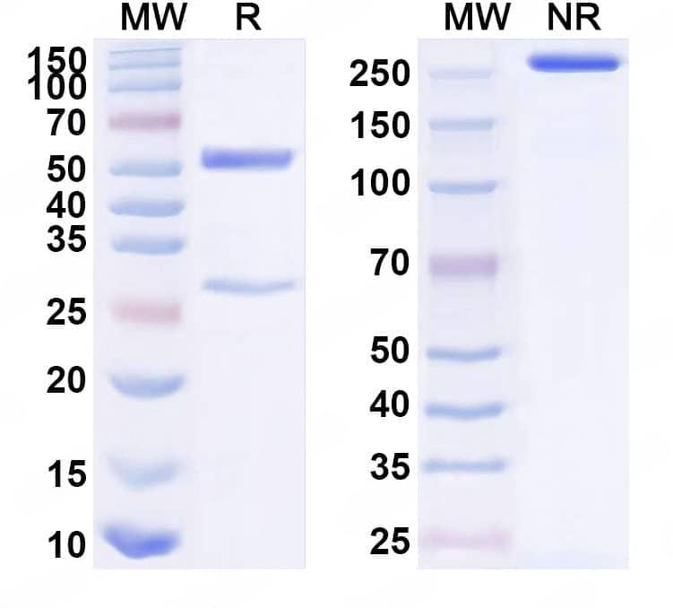
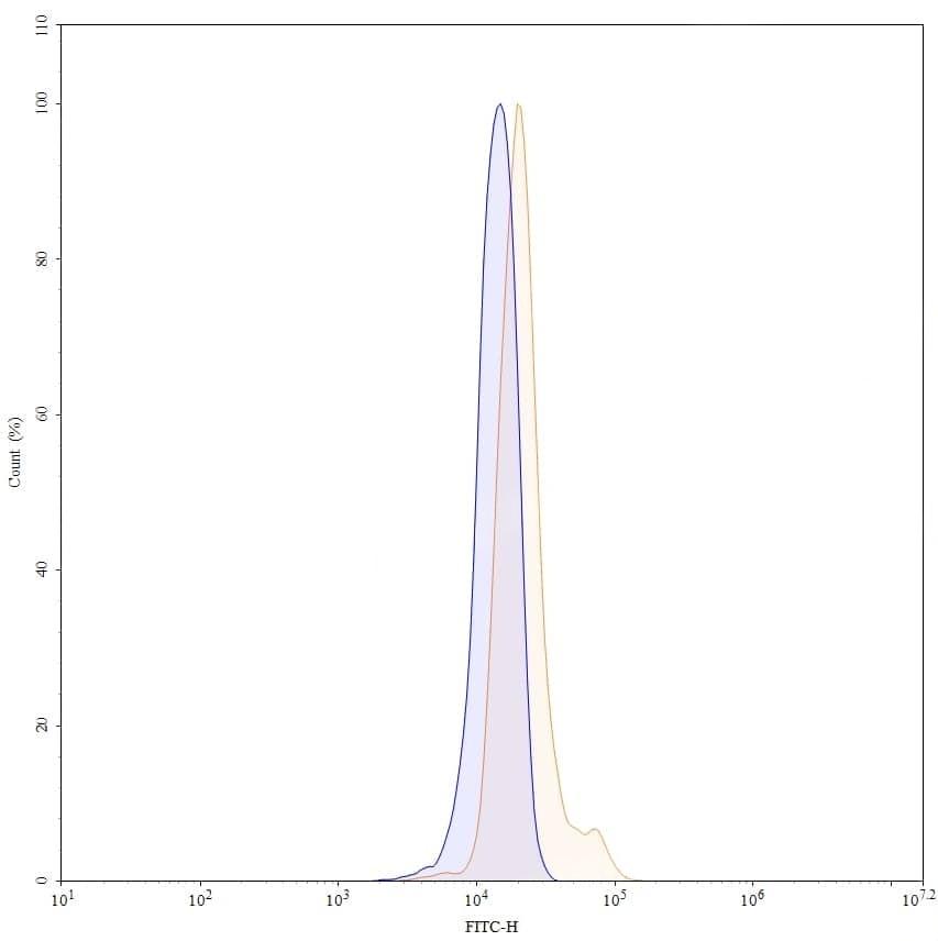
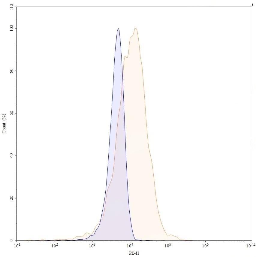
SEC-HPLC detection for Anti-Human BAX Antibody (6A7). | Flow-cytometry using anti-human BAX antibody.HepG2 cells were stained with an irrelevant antibody (Blue Histogram) or an anti-human BAX antibody monoclonal antibody (Catalog No.:TD-HC491013 ,Green Histogram) at a concentration of 5 µg/ml for 30 mins at RT. After washing, bound antibody was detected using a FITC conjugated goat anti-mouse antibody (Catalog No.:TD-MF690414) and cells analysed on a NovoCyte Flow Cytometer.,Flow-cytometry using anti-human BAX antibody.Human Jurkat cell line were stained with an irrelevant antibody (Blue Histogram) or an anti-human BAX antibody monoclonal antibody (Catalog No.:TD-HC491013 ,Green Histogram) at a concentration of 5 µg/ml for 30 mins at RT. After washing, bound antibody was detected using a FITC conjugated goat anti-mouse antibody (Catalog No.:TD-MF690414) and cells analysed on a NovoCyte Flow Cytometer.,Flow-cytometry using anti-human BAX antibody.A549 cells were stained with an irrelevant antibody (Blue Histogram) or an anti-human BAX antibody monoclonal antibody (Catalog No.:TD-HC491013 ,Green Histogram) at a concentration of 5 µg/ml for 30 mins at RT. After washing, bound antibody was detected using a FITC conjugated goat anti-mouse antibody (Catalog No.:TD-MF690414) and cells analysed on a NovoCyte Flow Cytometer. | SDS-PAGE for Anti-Human BAX Antibody (6A7). | Flow-cytometry using FITC anti-human BAX antibody. TNFa stimulated U937 cells were fixed and permeabilized, then stained with an irrelevant antibody (Blue Histogram) or an FITC anti-human BAX monoclonal antibody (Catalog No.:TD-HC491013, Yellow Histogram) at a concentration of 5 µg/ml for 30 mins at RT. After washing, and cells analysed on a NovoCyte Flow Cytometer.,Flow-cytometry using PE anti-human BAX antibody. TNFa stimulated U937 cells were fixed and permeabilized, then stained with an irrelevant antibody (Blue Histogram) or an PE anti-human BAX monoclonal antibody (Catalog No.:TD-HC491013, Yellow Histogram) at a concentration of 5 µg/ml for 30 mins at RT. After washing, and cells analysed on a NovoCyte Flow Cytometer.