用于亚细胞成像的荧光多肽
利用共焦显微镜或荧光显微镜开展体外成像,仍是研究细胞内各类生物学过程与相互作用的高效且有效的方法之一。当荧光标记多肽与该类成像技术相结合时,可精准识别细胞内的特定靶标,为亚细胞层面的观察与分析提供关键支持。
For example, cell penetrating peptides (CPPs) modified with FITC and photoactivatable probes have been used to track binding patterns and dynamic behavior over time. Unlike proteins, these peptides localize to specific targets on actin and are less prone to protein aggregation, making them ideal for in vitro tracking (Pan 2014). Similarly, FITC-labeled CPPs have been used to image intracellular compartments of cells with low risk of cytotoxicity (Kirkham 2015).
用于血管造影的荧光多肽
荧光标记多肽的主要应用领域之一是血管造影体内成像。该技术通过荧光标记多肽作为造影剂,对血管内部进行成像,帮助医生清晰掌握血管状况,进而为患者制定更适配的医疗策略与治疗方案。
For example, high resolution near-infrared fluorophores have been used as fibrin imaging-agents for deep vein thrombosis (DVT). Using Cy7 labeled fibrin-targeting peptides, CT scanning and confocal microscopy were used to differentiate between acute and subacute murine DVT (Hara 2012). Similar techniques have also been used for detecting apoptosis as a symptom for glaucoma. Fluorophore-labeled peptides that are activated by caspases can be imaged in vivo to track glaucoma progression (Qiu 2014).
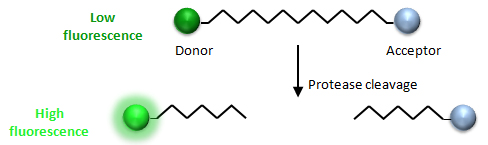
用于筛选蛋白水解肽的FRET
蛋白水解酶在传染性疾病中具有重要作用,因此成为新疗法开发的关键研究靶点。为识别这类酶所靶向的肽序列,肽文库是常用工具。其核心原理是将潜在的蛋白酶靶序列与 FRET 对相结合,当蛋白酶裂解靶肽时,即可检测到荧光信号,从而实现对蛋白水解肽的高效筛选。
In these studies, a donor molecule, such as Abz (Marcondes 2015) or Lucifer Yellow (Rossé 2000) is covalently attached to the C-terminus, while acceptor molecules (ex. Dabsyl, DNP) are coupled to the N-terminus. This strategy is commonly employed with peptides synthesized using a solid-phase approach, and peptides are left conjugated to the bead. If the peptides are not targeted by protease activity, both the donor and acceptor will be present, and the bead will appear non-fluorescent. If the peptides are cleaved, the peptide will no longer be quenched by FRET and the beads will fluoresce. Since protease studies are most effective when FRET is combined with shorter peptides, micro-scale peptide libraries are ideal choices for these experiments.