产品中心
Product Center
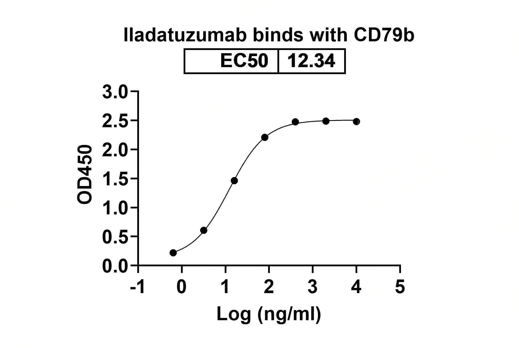
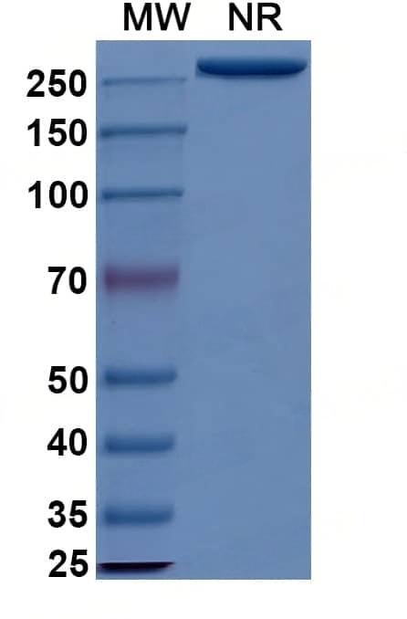
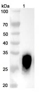
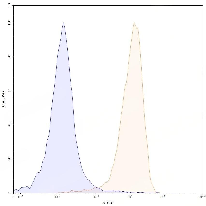
Research Grade Iladatuzumab
All
Bioactivity
SDS-PAGE
Western blot
- Catalog: TD-HW431026
- Clonality: Monoclonal
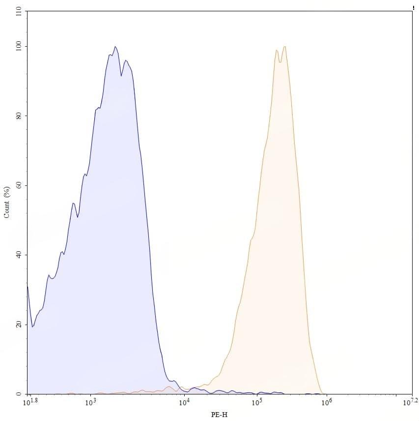
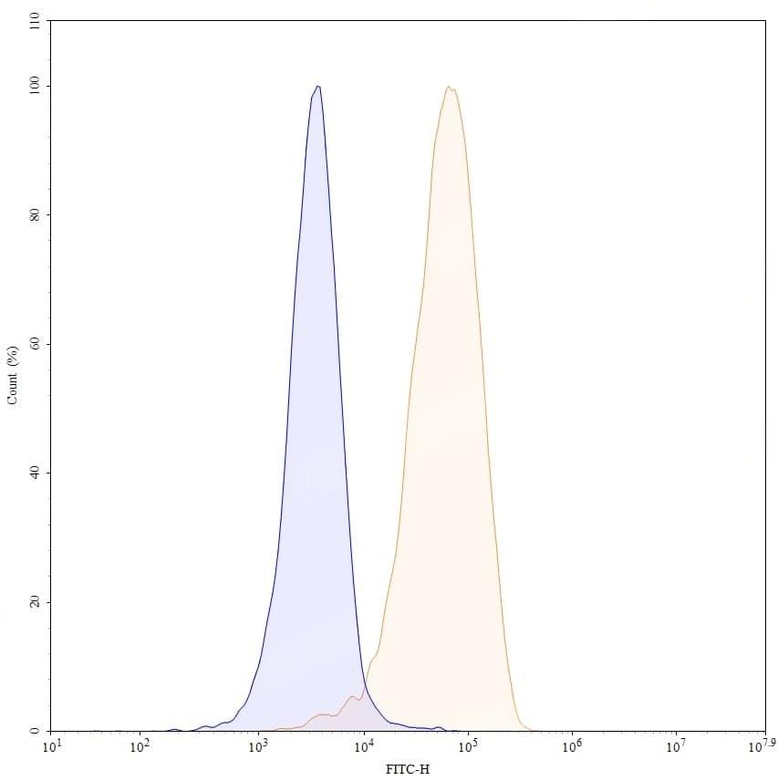
- Application: ELISA, Bioactivity: FACS, Functional assay, Research in vivo
- Synonyms: DCDS0780A, MCDS0593A, RO7032005, 1906205-76-2
-
规格:
询价
-
价格:
¥100ug