产品中心
Product Center
Mouse Monoclonal Antibody to CD1A
All

- Catalog: TD-30187
- Clonality: mAb
- Application: WB, IHC-P, ICC, ELISA
- Synonyms: R4; T6; CD1; FCB6; HTA1; CD1A
-
规格:
100ul 50ul
-
价格:
¥2180
Mouse Monoclonal Antibody to CD1A
|
|
| Description | |
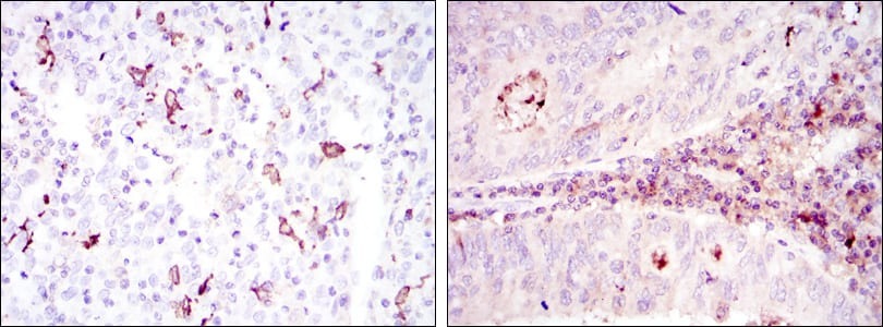
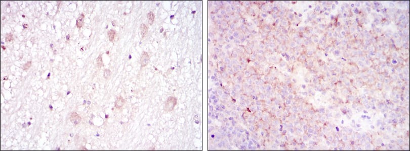
| CD1a is a non polymorphic MHC Class 1 related cell surface glycoprotein, expressed in association with Beta 2 microglobulin. CD1a is expressed by cortical thymocytes, Langerhan's cells and by interdigitating cells. CD1a is also expressed by some malignancies of T cell lineage and in histiocytosis X. Tissue specificity: Expressed on cortical thymocytes, epidermal Langerhans cells, dendritic cells, on certain T-cell leukemias, and in various other tissues. | |
| Specification | |
| Aliases | R4; T6; CD1; FCB6; HTA1; CD1A |
| Entrez GeneID | 909 |
| Swissprot | P06126 |
| clone | 7A7 |
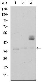
| WB Predicted band size | 37kDa |
| Host/Isotype | Mouse IgG1 |
| Antibody Type | Primary antibody |
| Storage | Store at 4°C short term. Aliquot and store at -20°C long term. Avoid freeze/thaw cycles. |
| Species Reactivity | Human |
| Immunogen | Purified recombinant fragment of human CD1A expressed in E. Coli. |
| Formulation | Ascitic fluid containing 0.03% sodium azide. |
| Application | |
| WB | 1/500 - 1/2000 |
| IHC | 1/200 - 1/1000 |
| ICC | 1/200 - 1/1000 |
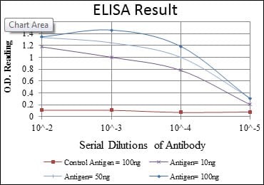
| ELISA | 1/10000 |
| Product Image | |