产品中心
Product Center
Mouse Monoclonal Antibody to CD31
All
- Catalog: TD-20131
- Clonality: mAb
- Application: IHC, IF
- Synonyms: CD31; PECAM-1; PECAM1
-
规格:
100ul 50ul
-
价格:
¥2180
Mouse Monoclonal Antibody to CD31
|
|
| Description | |
| CD31, also known as platelet endothelial cell adhesion molecule 1 (PECAM1), is a type I integral membrane glycoprotein and a member of the immunoglobulin superfamily of cell surface receptors.It is constitutively expressed on the surface of endothelial cells, and concentrated at the junction between them. The antibody reacts with the murine form of the Platelet-Endothelial Cell Adhesion Molecule. The reactivity of the antibody is restricted to the isoform of the molecule that is selectively expressed by endothelial cells.The antigen is predominantly present at the lateral borders of endothelial cells as described for human PECAM-1.It is also weakly expressed on many peripheral lymphoid cells and platelets.CD31 has been used to measure angiogenesis in association with tumor recurrence. Other studies have also indicated that CD31 and CD34 can be used as markers for myeloid progenitor cells and recognize different sub/sets of myeloid leukemia infiltrates (granular sarcomas). | |
| Specification | |
| Aliases | CD31; PECAM-1; PECAM1 |
| Entrez GeneID | 5175 |
| Swissprot | P16284 |
| clone | 2F7B2 |
| Host/Isotype | Mouse IgG1 |
| Antibody Type | Primary antibody |
| Storage | Store at 4°C short term. Aliquot and store at -20°C long term. Avoid freeze/thaw cycles. |
| Species Reactivity | Human |
| Immunogen | Purified recombinant fragment of human CD31 expressed in E. Coli. |
| Formulation | Ascitic fluid containing 0.03% sodium azide. |
| Application | |
| IHC | 1/200 - 1/1000 |
| IF | 1/200 - 1/1000 |
| ELISA | 1/10000 |
| Product Image | |
|
|
|
|
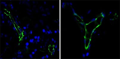
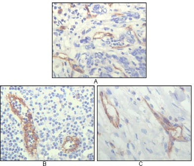
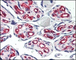
| Immunofluorescence analysis of paraffin-embedded human lung cancer(left) and breast cancer(right) cells using CD31 mouse mAb (green). Blue: DRAQ5 fluorescent DNA dye. | Immunohistochemical analysis of paraffin-embedded human lung cancer (A), lymphonodus tissue (B) and breast cancer (C), showing cytoplasmic localization of vascular endothelial cells using CD31 mouse mAb with DAB staining. | Immunohistochemical analysis of paraffin-embedded human lung cancer (A), lymphonodus tissue (B) and breast cancer (C), showing cytoplasmic localization of vascular endothelial cells using CD31 mouse mAb with DAB staining. |