产品中心
Product Center
Mouse Monoclonal Antibody to CD44
All

- Catalog: TD-20282
- Clonality: mAb
- Application: WB, IHC, IF, FCM
- Synonyms: IN; LHR; MC56; MDU2; MDU3; MIC4; Pgp1; CDW44; CSPG8; HCELL
-
规格:
100ul 50ul
-
价格:
¥2180
Mouse Monoclonal Antibody to CD44 |
|
| Description | |
| CD44, also known as IN, LHRMIC4, CDW44, HCELL. It is a cell-surface glycoprotein involved in cell-cell interactions, cell adhesion and migration. It is a receptor for hyaluronic acid (HA) and can also interact with other ligands, such as osteopontin, collagens, and matrix metalloproteinases (MMPs). This protein participates in a wide variety of cellular functions including lymphocyte activation, recirculation and homing, hematopoiesis, and tumor metastasis. Transcripts for this gene undergo complex alternative splicing that results in many functionally distinct isoforms, however, the full length nature of some of these variants has not been determined. Alternative splicing is the basis for the structural and functional diversity of this protein, and may be related to tumor metastasis. | |
| Specification | |
| Aliases | IN; LHR; MC56; MDU2; MDU3; MIC4; Pgp1; CDW44; CSPG8; HCELL |
| Entrez GeneID | 960 |
| Swissprot | P16070 |
| clone | 8E2F3 |
| WB Predicted band size | 82kDa |
| Host/Isotype | Mouse IgG1 |
| Antibody Type | Primary antibody |
| Storage | Store at 4°C short term. Aliquot and store at -20°C long term. Avoid freeze/thaw cycles. |
| Species Reactivity | Human,Mouse |
| Immunogen | Purified recombinant fragment of human CD44 (628-699) expressed in E. Coli. |
| Formulation | Ascitic fluid containing 0.03% sodium azide. |
| Application | |
| WB | 1/500 - 1/1000 |
| IHC | 1/200 - 1/1000 |
| IF | 1/200 - 1/1000 |
| FCM | 1/200 - 1/400 |
| ELISA | 1/10000 |
| Product Image | |
|
|
|
|
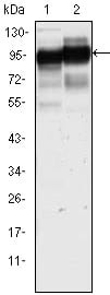
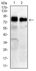
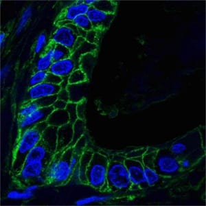
| Western blot analysis using CD44 mouse mAb against Hela (1) and HUVE-12(2) cell lysate. | Western blot analysis using CD44 mouse mAb against COS7(1),C6(2) cell lysate. | Confocal analysis of paraffin-embedded human lung cancer tissues using CD44 mouse mAb (green), showing membrane localization. Blue: DRAQ5 fluorescent DNA dye. |
|
|
|
|
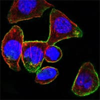
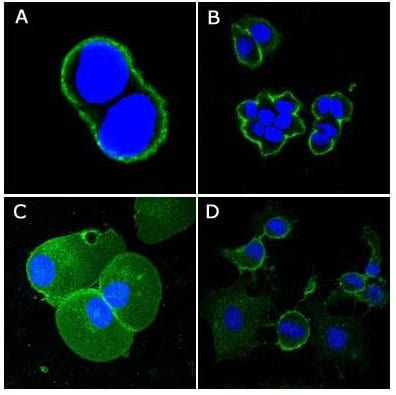
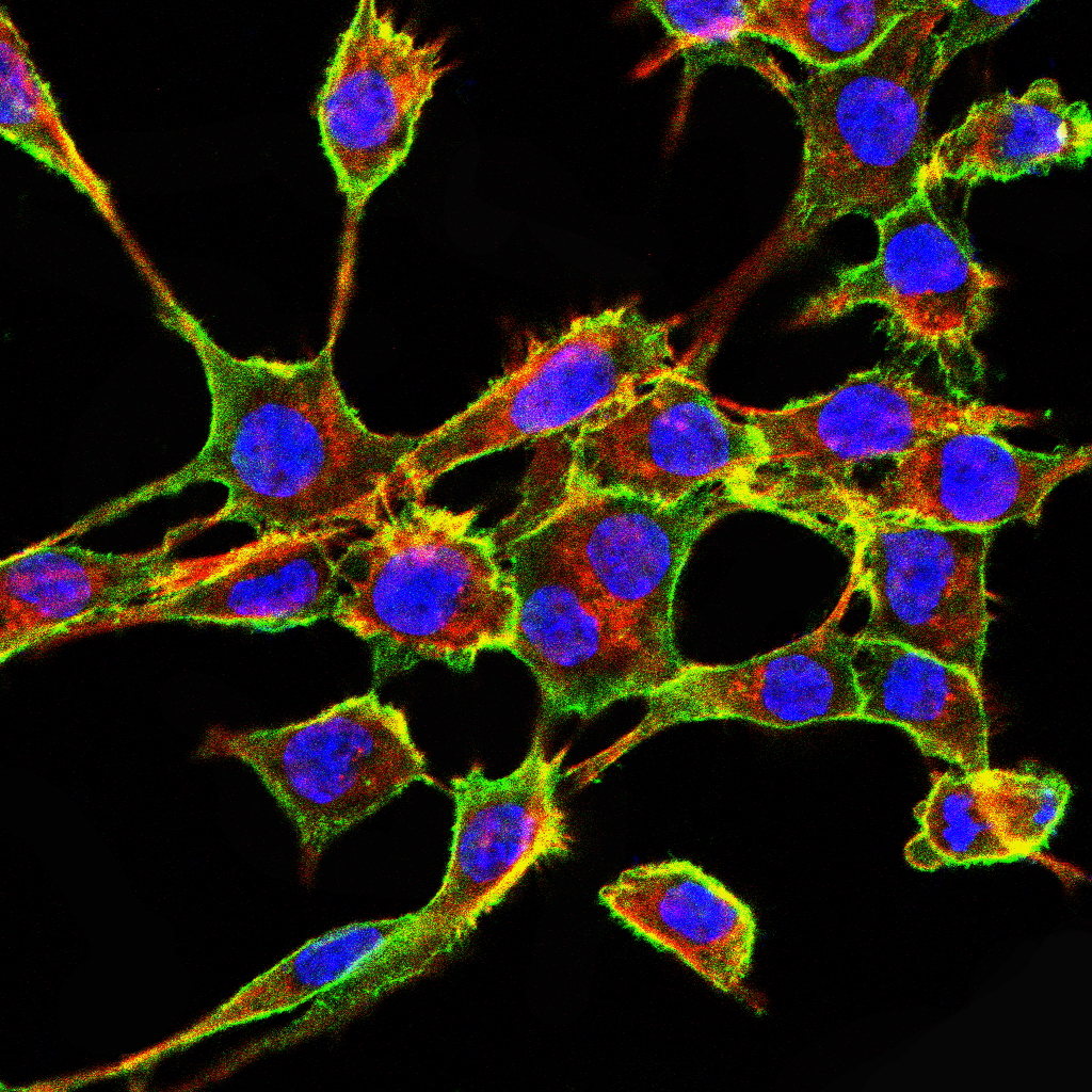
| Confocal Immunofluorescence analysis of PANC-1 cells using CD44 mouse mAb (green). Red: Actin filaments have been labeled with DY-554 phalloidin. Blue: DRAQ5 fluorescent DNA dye. | Confocal Immunofluorescence analysis of PANC-1 cells using CD44 mouse mAb (green). Red: Actin filaments have been labeled with DY-554 phalloidin. Blue: DRAQ5 fluorescent DNA dye. | Immunofluorescence analysis of RSC-96 cells using CD44 mouse mAb (green). Blue: DRAQ5 fluorescent DNA dye. Red: Actin filaments have been labeled with Alexa Fluor- 555 phalloidin. |
|
|
|
|
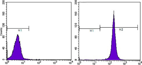
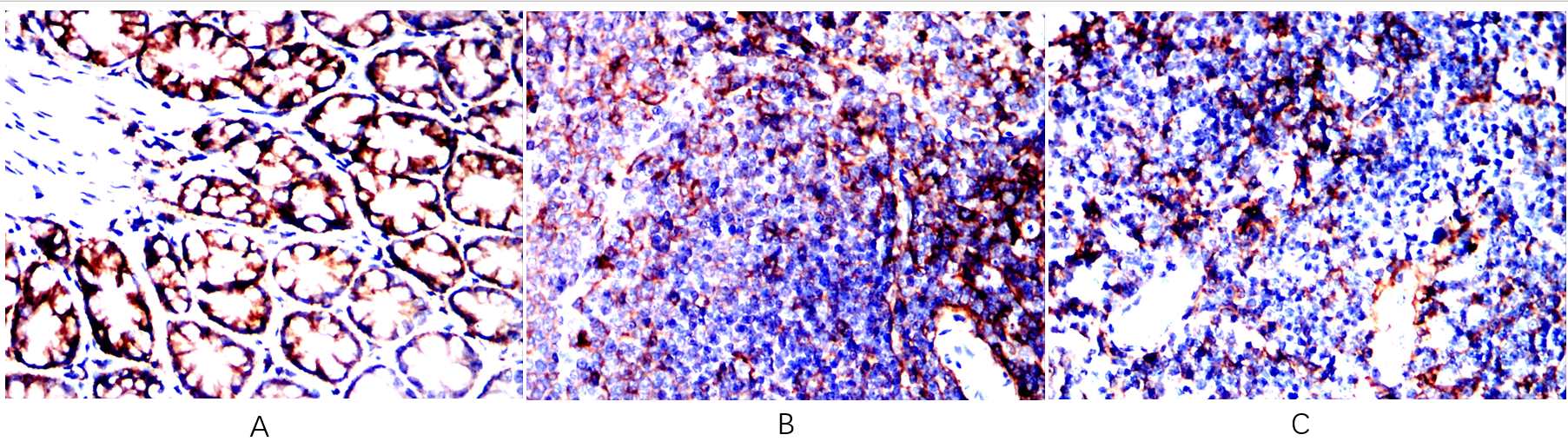
| Flow cytometric analysis of Hela cells using CD44 mouse mAb (right) and negative control (left). | Immunohistochemical analysis of paraffin-embedded human breast carcinoma tissues, showing membrane localization with DAB staining using CD44 mouse mAb. | Immunohistochemical analysis of paraffin-embedded Rat colon(A) Rat spleen(B) Rat thymus(C) using CD44 mouse mAb with DAB staining. |