产品中心
Product Center
CD126
All
ELISA
WB
IF
- Catalog: TD-32241
- Clonality: mAb
- Application: WB, IHC, IF, FCM
- Synonyms: IL6Q; gp80; CD126; HIES5; IL-6R; IL6RA; IL6RQ; IL-1Ra; IL-6RA; IL6QTL; IL-6R-1
-
规格:
100ul 50ul
-
价格:
¥2180
Mouse Monoclonal Antibody to CD126
| Description | |
| This gene encodes a subunit of the interleukin 6 (IL6) receptor complex. Interleukin 6 is a potent pleiotropic cytokine that regulates cell growth and differentiation and plays an important role in the immune response. The IL6 receptor is a protein complex consisting of this protein and interleukin 6 signal transducer (IL6ST/GP130/IL6-beta), a receptor subunit also shared by many other cytokines. Dysregulated production of IL6 and this receptor are implicated in the pathogenesis of many diseases, such as multiple myeloma, autoimmune diseases and prostate cancer. Alternatively spliced transcript variants encoding distinct isoforms have been identified in this gene. A pseudogene of this gene is found on chromosome 9. [provided by RefSeq, Aug 2020] | |
| Specification | |
| Aliases | IL6Q; gp80; CD126; HIES5; IL-6R; IL6RA; IL6RQ; IL-1Ra; IL-6RA; IL6QTL; IL-6R-1 |
| Entrez GeneID | 3570 |
| Swissprot | P08887 |
| clone | 8F7G6 |
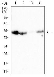
| WB Predicted band size | 52kDa |
| Host/Isotype | Mouse IgG1 |
| Antibody Type | Primary antibody |
| Storage | Store at 4°C short term. Aliquot and store at -20°C long term. Avoid freeze/thaw cycles. |
| Species Reactivity | Human, Mouse |
| Immunogen | Purified recombinant fragment of human CD126 (AA: EXTRA 20-177) expressed in E. Coli. |
| Formulation | Purified antibody in PBS with 0.05% sodium azide |
| Application | |
| WB | 1/500 - 1/2000 |
| IHC | 1/200 - 1/1000 |
| IF | 1/200 - 1/1000 |
| FCM | 1/200 - 1/400 |
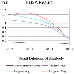
| ELISA | 1/10000 |
| Product Image | |
|
Black line: Control Antigen (100 ng);Purple line: Antigen (10ng); Blue line: Antigen (50 ng); Red line:Antigen (100 ng)
|
Western blot analysis using CD126 mouse mAb against Jurkat (1), MOLT4 (2), Raw264.7 (3)and THP-1 (4) cell lysate.
|
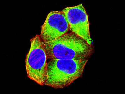
Immunofluorescence analysis of Hela cells using CD126 mouse mAb (green). Blue: DRAQ5 fluorescent DNA dye. Red: Actin filaments have been labeled with Alexa Fluor- 555 phalloidin. Secondary antibody from Fisher (Cat#: 35503) |
|
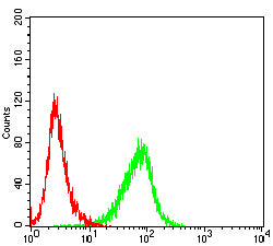
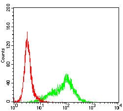
Flow cytometric analysis of Jurkat cells using CD126 mouse mAb (green) and negative control (red).
|
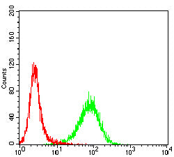
Flow cytometric analysis of K562 cells using CD126 mouse mAb (green) and negative control (red).
|
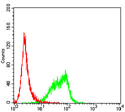
Flow cytometric analysis of THP-1 cells using CD126 mouse mAb (green) and negative control (red).
|
|
Flow cytometric analysis of U937 cells using CD126 mouse mAb (green) and negative control (red).
|
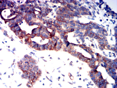
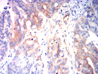
Immunohistochemical analysis of paraffin-embedded human ovarian cancer tissues using CD126 mouse mAb with DAB staining.
|
Immunohistochemical analysis of paraffin-embedded human rectal cancer tissues using CD126 mouse mAb with DAB staining.
|