Caption
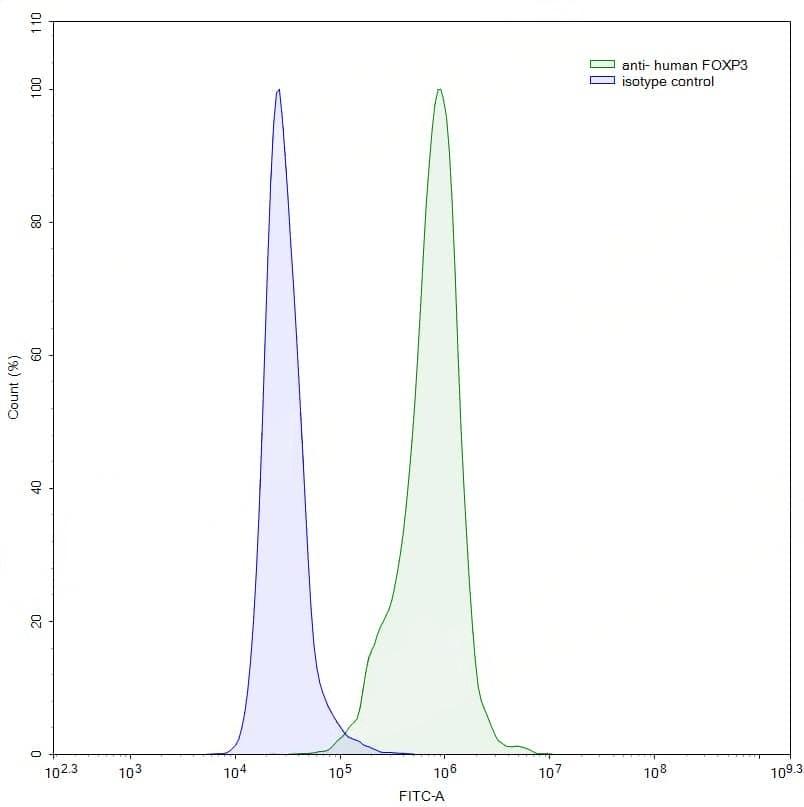
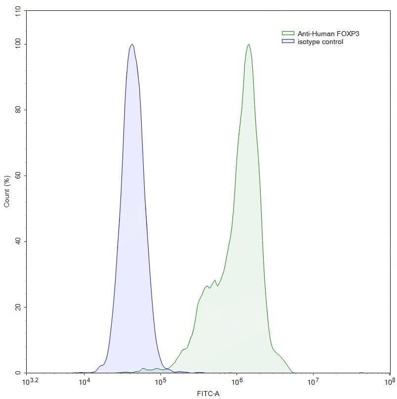
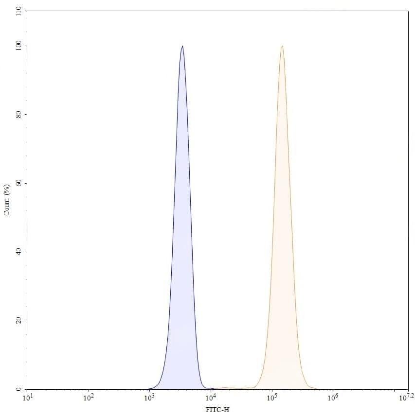
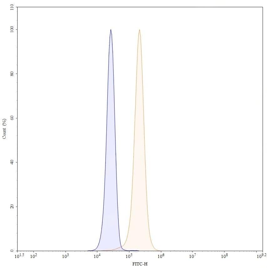
Flow-cytometry using anti-human FOXP3 antibody. Jurkat cells were stained with an irrelevant antibody (Blue Histogram) or an anti-human FOXP3 antibody monoclonal antibody (Catalog No.:TD-HV388025, Green Histogram) at a concentration of 5 µg/ml for 30 mins at RT. After washing, bound antibody was detected using a FITC conjugated goat anti-mouse antibody (TD-MF690414) and cells analysed on a NovoCyte Flow Cytometer.,Flow-cytometry using anti-human FOXP3 antibody. Hela cells were stained with an irrelevant antibody (Blue Histogram) or an anti-human FOXP3 antibody monoclonal antibody (Catalog No.:TD-HV388025, Green Histogram) at a concentration of 5 µg/ml for 30 mins at RT. After washing, bound antibody was detected using a FITC conjugated goat anti-mouse antibody (TD-MF690414) and cells analysed on a NovoCyte Flow Cytometer. | Flow-cytometry using anti-human FOXP3 antibody. Jurkat cells were fixed and permeabilized, then stained with an irrelevant antibody (Blue Histogram) or an anti-human FOXP3 monoclonal antibody (Catalog No.:TD-HV388025, Yellow Histogram) at a concentration of 5 µg/ml for 30 mins at RT. After washing, bound antibody was detected using Goat Anti-Mouse IgG H&L Polyclonal Antibody, FITC (Catalog No.:TD-MF690414) and cells analysed on a NovoCyte Flow Cytometer.,Flow-cytometry using anti-human FOXP3 antibody. Hela cells were fixed and permeabilized, then stained with an irrelevant antibody (Blue Histogram) or an anti-human FOXP3 monoclonal antibody (Catalog No.:TD-HV388025, Yellow Histogram) at a concentration of 5 µg/ml for 30 mins at RT. After washing, bound antibody was detected using Goat Anti-Mouse IgG H&L Polyclonal Antibody, FITC (Catalog No.:TD-MF690414) and cells analysed on a NovoCyte Flow Cytometer.