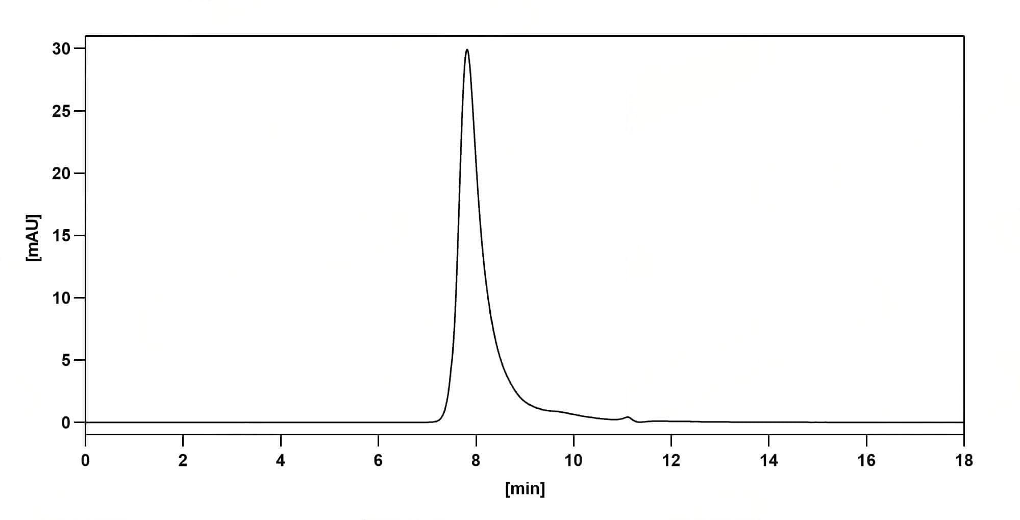
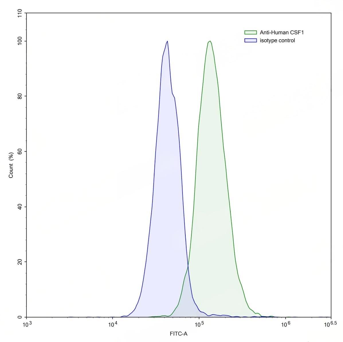
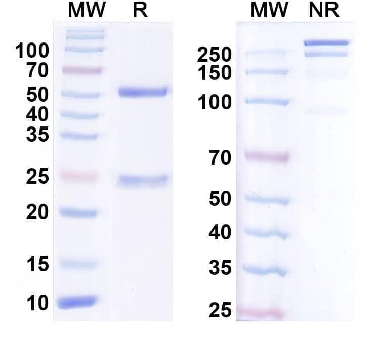
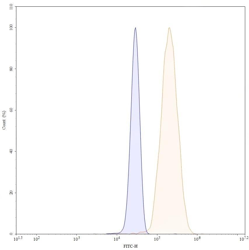
Caption
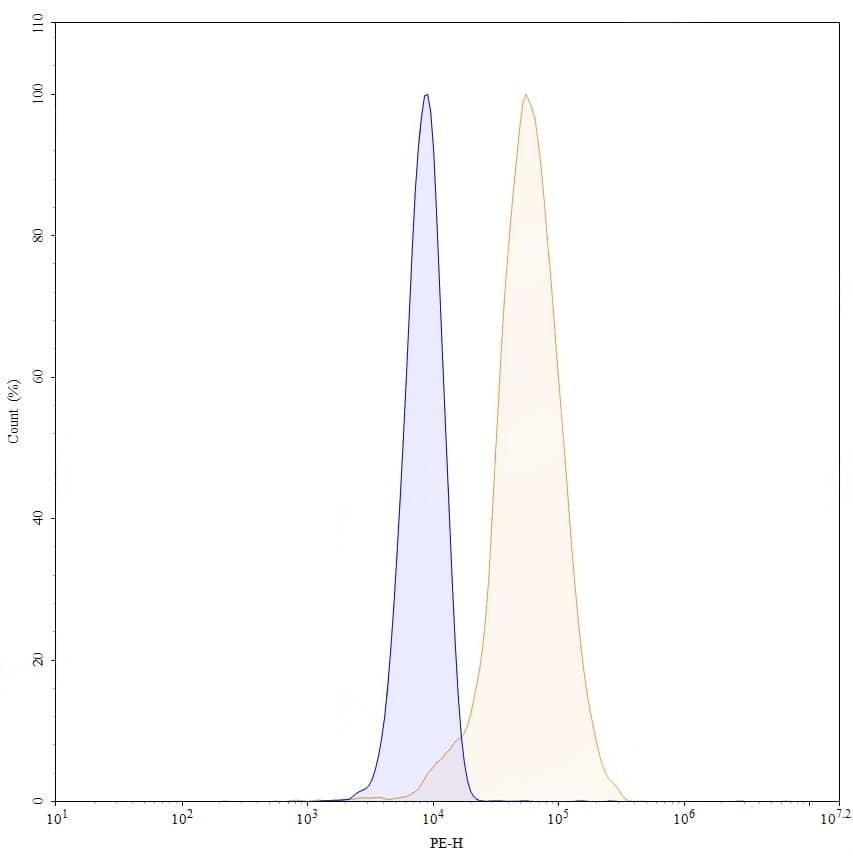
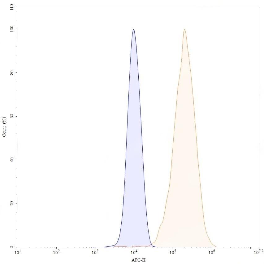
SEC-HPLC detection for InVivoMAb Anti-Human CSF1/M-CSF (ABV0114). | Flow-cytometry using anti-human CSF1 antibody.MG-63 cells were stained with an irrelevant antibody (Blue Histogram) or an anti-human CSF1 antibody monoclonal antibody (Catalog No.:TD-HY515010 ,Green Histogram) at a concentration of 5 µg/ml for 30 mins at RT. After washing, bound antibody was detected using a FITC conjugated goat anti-human antibody (Catalog No.:TD-HF690414) and cells analysed on a NovoCyte Flow Cytometer. | SDS-PAGE for InVivoMAb Anti-Human CSF1/M-CSF (Iv0052). | Flow-cytometry using FITC anti-human CSF1 antibody. MG-63 cells were fixed and permeabilized, then stained with an irrelevant antibody (Blue Histogram) or an FITC anti-human CSF1 monoclonal antibody (Catalog No.:TD-HY515010, Yellow Histogram) at a concentration of 5 µg/ml for 30 mins at RT. After washing, cells analysed on a NovoCyte Flow Cytometer.,Flow-cytometry using PE anti-human CSF1 antibody. MG-63 cells were fixed and permeabilized, then stained with an irrelevant antibody (Blue Histogram) or an PE anti-human CSF1 monoclonal antibody (Catalog No.:TD-HY515010, Yellow Histogram) at a concentration of 5 µg/ml for 30 mins at RT. After washing, cells analysed on a NovoCyte Flow Cytometer.,Flow-cytometry using APC anti-human CSF1 antibody. MG-63 cells were fixed and permeabilized, then stained with an irrelevant antibody (Blue Histogram) or an APC anti-human CSF1 monoclonal antibody (Catalog No.:TD-HY515010, Yellow Histogram) at a concentration of 5 µg/ml for 30 mins at RT. After washing, cells analysed on a NovoCyte Flow Cytometer.